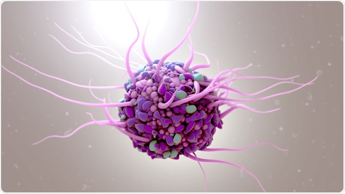
The cellular microenvironment is defined as the local environment surrounding a cell, which contains physical and chemical signals that can influence cellular behavior, either directly or indirectly.
 Design_Cells | Shutterstock
What is the cellular microenvironment composed of?
A cell’s microenvironment includes the extracellular matrix; similar or dissimilar cells that surround another cell; different cytokines, hormones, and reactive species; local physical properties of a cell; the mechanical forces that are produced by the movement of molecular motors or fluids inside a cell.
The importance of each factor depends on the nature of the cell or tissue, and on the combination of these factors which ultimately will influence the behavior of the cells/tissue in the microenvironment.
Dynamic nature of the cellular microenvironment
The extracellular matrix is very heterogeneous and dynamic and also undergoes dynamic remodeling in space and time. The nature of the interaction between the microenvironment and cells is bidirectional. This means that as cells breakdown, they produce, rearrange and realign the components of extracellular matrix. Similarly, the matrix itself can regulate and modify the activity of cells. A detailed understanding of the nature of these interactions is integral for engineering tissues or organs.
How does the cellular microenvironment influence behavior?
Various aspects of microenvironment can influence cell behavior. In one study by Satyam and co., it was shown that the local accumulation of macromolecules can lead to improved secretion of extracellular matrix proteins from corneal fibroblasts.
In another study by Fujie and co., it was shown that micropatterned membranes can be used to develop retinal pigment epithelial layers, which can be implanted into the eye with minimal invasion. Also, mechanically confining cells in a microfluidic platform has been shown to regulate the process of mitosis.
Mimicking the cellular microenvironment
The cells in the body do not exist in isolation. They are surrounded by other cells and tissues that provide topographical and biochemical signals. The physical structures include nanopores in capillaries, nanofibers in the basement membrane, and nanocrystals in the bone microstructure.
Cell alignment is another factor that has been shown to be critical for muscle, blood vessel, nerve, and corneal architecture and functioning. In many cases, methods have been developed to induce cell alignment and the formation of a topographic pattern.
Using microfabrication and nanofabrication, complex surface features can be created and various patterns of shape, periodicity, and dimensions can now be reproduced. Similarly, construction of patterned substrates has also become a critical tool while created engineered tissues.
Patterned surfaces have been used for directing growth and differentiation processes. In one study, the use of nanopillars and increasing their height from 35nm to 400nm lead to an increased yield of neurons. Similarly, topographical cues have been shown to increase the expression of neuronal markers.
Can you modify the cellular microenvironment?
Microenvironment can also be tweaked to produce a certain outcome. For example, the microenvironment surrounding stem cells is a growing field of research, where the modification of the microenvironment can lead to an increase in cell numbers. This method can be used in the field of tissue engineering and regenerative medicine.
There are several stem cell reservoirs in the body and the microenvironment determines the plasticity and quiescence of these cells. Thus, the body keeps these cells in a state of quiescence or activity based on need and this switch is often regulated by the components of the microenvironment. Also, failure to regulate this switch may lead to de-differentiation or tumors.
Controlling the cellular microenvironment using microfluidics
Microfluidics is an emerging field where the fluids can be manipulated and controlled using networks that range in microliter to picoliter size. Manipulating the fluid flow has been used as a cue as a chemical and mechanical cue that can affect the cellular microenvironment in a three-dimensional space.
Further Reading