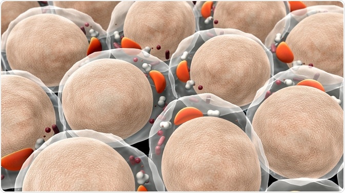
A fundamental aspect of biology is the study of cell structure and development. Recently, new methods have been developed to study nanoscale structures within the cell such as nucleosomes, actin and microtubules.
 Credit: URGREEN 3S/Shutterstock.com
Defining cell architecture at the mesoscale (i.e. at length scales that are larger than the molecular machinery) increases knowledge of cell function.
However, many subcellular biological structures lie below the diffraction limit of visible light, meaning conventional light microscopy cannot be utilized for visualizing all cell architecture.
Point-localization super-resolution imaging
Point-localization super-resolution imaging is a type of super-resolution (SR) imaging that achieves high resolution by uniting fluorescent labeling that is molecule specific with nanoscale spatial resolution. The method works by producing individual images through the isolation of neighboring molecules with their own fluorescence emission.
The individual images provide visualization of cell structures separated by at least 200 nanometers. A super-resolution image is developed by fitting the images through a point spread function.
The computer algorithm then calculates the centroid of the fluorescent light emission and therefore localizes the molecule. The process is repeated multiple times with each localized molecule represented as a point in the final image.
Three factors affect the spatial resolution produced in point-localization super-resolution. The optical resolution is dependent on the number of photons detected from a fluorescent molecule, and therefore the type probe molecule affects the image resolution. Sampling density is another important factor as the maximum spatial resolution is limited to twice the sampling density.
Additionally, increasing probe sizes also reduces the resolution. For some methods the dyes used have to be connected to a targeting molecule, usually antibodies, which restrict the visualization of cell architecture.
Soft X-ray tomography and correlated cryo-light microscopy
Though fluorescence-based imaging can determine the location and quantity of subcellular molecules at low concentrations, alternative techniques have been introduced to study reactions between structures.
Soft X-ray tomography and correlated cryo-light microscopy can be combined to define the molecules that are interacting, where the reactions are taking place, and the consequences for the cell. Correlated cryo-light microscopy is applied first to collect fluorescence images at low temperatures.
In the new generation of cryogenic microscopes, the sample is immersed in organic fluid such as liquid propane at a low cryogenic temperature. The use of organic immersion fluids means that high numerical aperture optics can be applied, allowing for improved magnification.
The sample is not fixed or stained providing the advantage of imaging the cell structures within its native environment. The location of mesoscale molecules is subsequently found by fluorescence microscopy.
To increase the detail of the cellular architecture being imaged, soft X-ray tomography is utilized. The method works through the principle of imaging cells by employing X-rays as the source of illumination.
Soft X-ray tomography images samples with high contrast because highly solvated regions of the cell appear transparent in comparison to the opaque areas produced from the regions with high molecule density.
The cell image can be segmented further depending on the linear absorption contrast (LAC) unit of each region in the cell. LACs are correlated with biomolecule density so the cell structure can be delineated and segmented – depending on whether the region is composed of dense biomolecules such as lipid droplets or low biomolecule density such as vacuoles. The segments can be used to study the internal structure of organelles.
Inside a cell
Advantages of the combined methods for visualizing cell architecture
A main advantage of soft X-ray tomography is that it does not require damaging contrast-enhancing stains such as platinum. While X-rays produce two-dimensional images, they can be easily transferred to three-dimensions by simply collecting a series of images at regular increments around a rotation axis.
The serialized application of the two discrete imaging methods, soft X-ray tomography and correlated cryo-light microscopy, provides three-dimensional views of the cell architecture by combining high-resolution structural information with localization data of individual molecules.
Sources:
- Mistelli, T. 2001 The concept of self-organization in cellular architecture, Journal of Cell Biology, 155, pp. 181-186.
- Sengupta, P. et al. 2012. Visualizing Cell Structure and Function with Point-Localization Superresolution Imaging, Developmental Cell, 23, pp. 1092-1102.
- McDermott, G. et al. 2012. Visualizing Cell Architecture and Molecular Location Using Soft X-Ray Tomography and Correlated Cryo-Light Microscopy, Annual Review in Physical Chemistry, 5, pp. 225-239.
- Ekman, A.A. et al. 2017. Mesoscale imaging with cryo-light and X-rays: Larger than molecular machines, smaller than a cell, Biology of the Cell, 109, pp. 24-38.
Further Reading