Flow cytometry is a rapid method of cell counting and sorting, in some cases incorporating several characterization techniques such as mass spectrometry and fluorescence microscopy that can allow greater analysis to be undertaken in situ.
During flow cytometry cells are passed in single file to be analyzed individually, usually then categorized for further examination by other methods. Cell size, optical density, morphology, granularity, and several other physical parameters that can be determined by light scattering can be used to infer the health of the cell, but the value of flow cytometry is significantly improved and expanded when incorporating other characterization techniques, amongst which the use of fluorophores in conjunction with fluorescence microscopy has proven extremely useful.

Image Credit: Jarun Ontakrai/Shutterstock.com
Biomarker identification
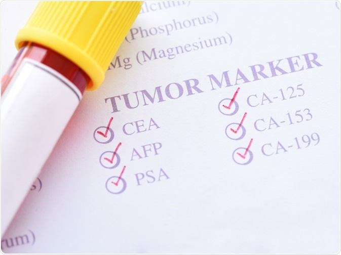
Biomarkers are molecules that can be used to quantitatively indicate the type and stage of a disease in a patient, allowing physicians to choose the best course of treatment. For example, the expression of estrogen receptor-alpha is a favorable prognostic factor in breast cancer patients, suggesting that patients will respond well to hormone therapy.
Biomarker discovery usually takes either a proteomic, metabolomic, lipidomic, or glycomic approach, focusing on analyzing the expression of proteins, all metabolites, lipids, or glycans, respectively, to uncover relationships between them and disease.
Several analytical methods are utilized in biomarker detection, involving collecting or culturing diseased cells or tissues and subsequently assessing the cell population for common abnormalities. Enzyme-linked immunosorbent assays are frequently used for this purpose, with the protein or other biomolecules of interest being tagged and allowing them to be quantitatively identified. However, this method is not able to distinguish between biomarkers produced by individual cells, while flow cytometry is capable of tracking multiple physical and biochemical parameters simultaneously in each cell.
Flow cytometry is extensively employed in cell sorting, allowing cells bearing features of interest to be separated and analyzed in greater detail, often undergoing detailed microscopy at the identification stage of biomarker discovery to determine the mechanism of disease. Researchers also employ flow cytometry to gather together rare cells that would otherwise be difficult to collect in abundance, such as stem cells.
Flow cytometry in diagnosis and prognosis
A variety of sample types can be utilized in flow cytometry, including whole blood, cell lines, tissues, and frozen peripheral blood cells, though the former is usually preferred to better represent the cell population in the blood.
Further Reading