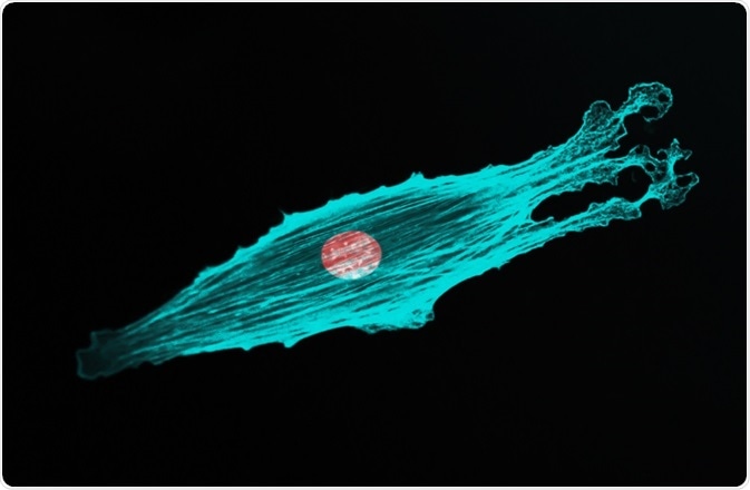
Fluorescence microscopy is now an essential part of cell biology. Developments to this imaging technique have improved the visualization and data gathered using fluorescence microscopy.

DrimaFilm | Shutterstock
What is fluorescence microscopy?
Fluorescence microscopy is an optical microscopy technique that utilizes fluorescence and phosphorescence to study the properties of organic or inorganic substances. Fluorescence is mainly used in cell biology, but it is also utilized in other bioscience fields, as well as in material sciences.
In the life sciences, researchers carrying out fluorescence microscopy typially use a technique known as epifluorescence microscopy. This involves the light of the required excitation passing through an excitation filter, and then an objective lens where it can illuminate the subject.
The fluorescence is emitted from the subject and focused through the same objective that the original light source passed. After passing the objective lens, the emitted fluorescence travels through an emission filter into an ocular lens and is focused onto the detector.
There are four main light sources used during fluorescence microscopy:
- Xenon arc lamps or mercury-vapor lamps
- Lasers
- Supercontinuum sources
- High power light-emitting diodes (LEDs)
When disadvantages of using fluorescence microscopy are discussed, it must be emphasized that the electrons in the subject excited by the fluorescence cause chemical damage which results in photobleaching. Photobleaching causes fluorophores to lose their ability to fluoresce, which limits the time a sample can be observed by fluorescent microscopy.
A brief history of fluorescence microscopy
Fluorescence was first discovered in 1845 by Fredrick W. Herschel. He discovered that UV light can excite a quinine solution (e.g. tonic water) to emit blue light. British scientist Sir George G. Stokes further studied this discovery, and he observed that fluorescence emission from an object represents a longer wavelength than the UV light that originally excited the object.
Many decades later, in the early 1900s, the first uses of fluorophores in biological investigations were performed to stain tissues, bacteria, and other pathogens. This was later developed into fluorescence microscopy by Carl Zeisi and Carl Reichert.
Fluorescence labeling was achieved by Ellinger and Hirt in the early 1940s. The cloning of green fluorescent protein (GFP) was achieved in the early 1990s and was easily applied to fluorescence microscopy.
Advancing the fluorescence microscopy technique
As a result of many research studies examining the excitation and emission properties of substances, fluorescence microscopy can now be applied in a very controlled manner, providing extremely detailed results.
To improve the resolution of images gained during the early uses of fluorescence microscopy, pinhole apertures were introduced in the beam path of the microscope set-up. The set up involves placing one pinhole in front of the light source and another in front of the detector.
Both holes focus the emission light and the excitation light, respectively. This method is called the ‘double focusing system’ and was the basis for modern confocal microscopes.
Spinning disk microscopy is a confocal microscopy variant which was developed to reduce the image acquisition time and the exposure of the sample to laser light. The microscope passes the laser beam through a rotating disk that contains multiple pinhole apertures, allowing many spots on the specimen to be illuminated simultaneously to provide higher frame rates.
Another fluorescence microscopy variant that has been developed is the ‘f-photon microscopy’ technique, which involves the absorption of two photons simultaneously instead of one. This technique causes the excitation wavelength to be longer than the emission wavelength.
Finally, there is no out of focus light generated when using two-photon microscopy, meaning there is less photobleaching and photo-damage.
Further Reading