Skip to
The cell nucleus is the site of many important biological functions of the eukaryotic cell. These processes include transcription, replication, splicing and ribosome biogenesis. The effect of these processes extends to affecting cellular metabolism and growth. The nucleus contains approximately 2m of DNA which is enmeshed by the nuclear envelope, a crosslinked network of proteins and membranes.
The nucleus is mechanically stable, possessing the ability to resist deformation. Additionally, the nucleus dynamically interacts with the surrounding cytoskeleton. The mechanical support and functional organization of the nucleus is contributed by several nuclear subcompartments, or nuclear bodies. These facilitate the various nuclear processes, particularly gene expression. These subcompartments together form the nucleoskeleton, and include the following:

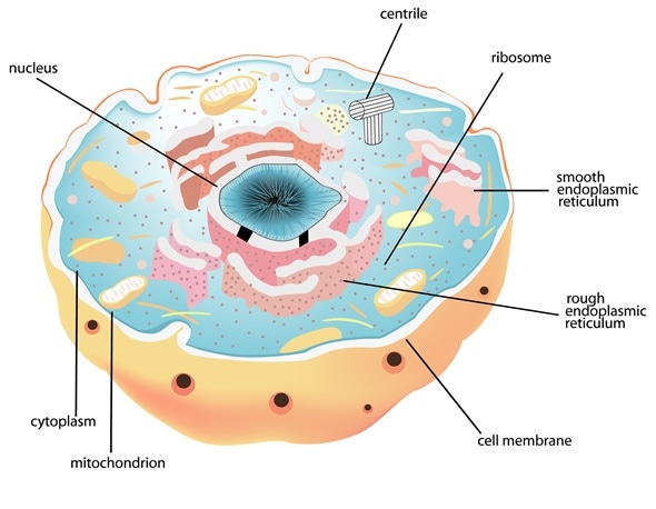
Cell and Nucleus, Image Copyright: sanjayart / Shutterstock
Nuclear Membrane (nuclear envelope)
The Nuclear Lamina is a structure that is located near the inner nuclear membrane. It is highly proteinaceous and is fibrillar in appearance. The fibrillar nature of the lamina arises from the building blocks of the lamina; the laminin proteins. These first form dimers, and subsequently orientate in a head-to-tail fashion to produce filaments.
The nuclear skeletal network (karyoskeleton) that results serves as an anchor for chromatin. This maintains the shape of the nucleus, spaces the nuclear pore complexes and organizes heterochromatin, the condensed region of DNA of nucleus. Additionally, it functions in nuclear processes such as DNA replication, particularly in the regulation of transcription factors.
The nuclear envelope itself has both an inner and outer nuclear lipid membranes. The outer nuclear membrane is continuous with the rough endoplasmic reticulum. Intermediate filaments form a network the nuclear lamina whilst other other inner nuclear membrane proteins mediate the interactions of the envelope with chromatin.
Nucleoskeleton
This is a network that provides support to the whole nucleus. It is made up of intermediate V type filaments composed of lamin proteins. It forms an elastic structure near the nuclear periphery and affords a viscoelastic property to the nucleus. At the nuclear envelope, the A and C type laminins provide a stiffness to the otherwise unstructured nucleus, whilst simultaneously supporting DNA processing events such as chromatin remodeling during replication.
B-type laminins provide support the processes of transcription and cellular signaling. There are also other structural proteins in the nucleoskeleton which impact the spatial organization of lamin proteins and other mechanical properties, such as resilience in response to stretch.
Nuclear Pore (Nuclear Pore Complex)
Nuclear pores are large protein complexes that form openings in the nuclear membrane. They sit at the point at which the inner nuclear membrane fuses with the outer nuclear membrane. They form aqueous channels that allow the selective movement of proteins, mRNA, tRNA, ribosome subunits and viruses in a bidirectional manner.
Molecules smaller than ∼40 kDa can pass freely across the nuclear envelope and do not require passage through the NPC. Molecules >40 kDa each have a specific means of transportation across the nuclear envelope. Collectively, the system of transport is comprised of the protein cargo, nuclear transport receptors (NTRs) such as importins and exportins, and the small GTPase Ran.
The NTRs are regulated by Ran; it mediates the association of the NTRs with the cargo and provides the energy which drive the transport across the membrane. It is estimated that a typical mammalian cell has between 3000 and 4000 nuclear pores on its nuclear membrane and these pores have a diameter of approximately 600 A°.
Nucleolus
Nucleoli are small spherical bodies that are located in the nucleus; they are usually found in a centralized region but may be found close to the nuclear membrane. Nucleoli are distinct from the nuclear material, being built by the nucleolus organizing region (NOR) of a chromosome. The NORs store the genes required for full ribosomal synthesis - these genes specifically encode ribosomal RNA subunits.
There are approximately 10 NORs per nucleolus, and several chromosomes collaborate in nucleolus assembly, so that at most, cells contain an average of one or two. The presence of the nucleoli is determined by the cell identity; most animal and plant cells have a nucleolus. However, the presence of nucleoli is usually indicative of malfunction; for example, malignant, ageing or starving cells frequently display nucleoli of varying size, abundance and shape.

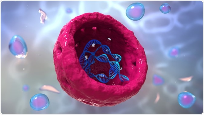
Nucleolus, human body cell 3d illustration. Image Credit: urfin / Shutterstock
In addition to DNA, the nucleolus is composed of two main regions – the pars fibrosa and the pars granulosa. The pars fibrosa contains proteins that participate in transcription and the pars granulosa houses the ribosomal precursors. Within the pars fibrosa, there is a fibrillar center which is distinct from a dense fibrillar component of the pars fibrosa.
The ribosomal RNA (rRNA) genes undergo transcription at the boundary of the two regions. Pre-rRNA processing follows this in the dense fibrillar component and continues in the granular component, where assembly of the rRNA with ribosomal proteins forms partially completed pre-ribosomal subunits ready for export to the cytoplasm.
Aside from its function in ribosome biogenesis, the nucleolus is also the site of processing of several other noncoding RNAs. The nucleolus additionally plays a role in controlling the stability of the protein p53, a critical cell-cycle regulator.
The nucleolus disassembles during the prophase stage of mitosis; however, the dense fibrillar component remains associated with chromosomes and forms a secondary constriction point on the chromosome called the nucleolus-organizing region (NOR). This aids in genome organization during the subsequent interphase stage of the cell cycle.
Chromosomes
Chromatin is a superstructure formed by highly organized compaction of the cell’s DNA and associated proteins. Histone proteins are the composite piece of the basic unit of chromatin organization called the nucleosome. Histones are basic proteins, possessing a positive charge which enables them to bind the negatively charged phosphate backbone of DNA.
Within the histone family, four histone members form an octamer which defines the nucleosomal protein component. Two H3 and two H4 proteins first tetramerize and combine with two H2A/H2B dimers. Approximately 150bp length of DNA wrap around each of the disk-shaped protein structure for approximately 2 turns.
The resultant DNA-histone complex forms the nucleosome core particle (NCP). Between each NCP is a region termed the linker region; this is comprised of between 10-90 bp of DNA together with the Histone subtype H1. Together, the linker DNA and NCP comprise the nucleosome; it repeats approximately every 200 bp. This produces a beads-on-a-string structure. This most basic unit of DNA can be further organized into a higher-order structure called chromatin. This is a 30nm fibre that forms from coiling of the nucleosomes into a solenoid.
Further still, the chromatin can coil even further to form the resultant chromosome. In the metaphase stage of the cell cycle, hypercondensation of the chromosome occurs, to allow correct segregation of the chromosomes in both meiosis and mitosis.
Further Reading