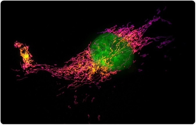
Stochastic Optical Reconstruction Microscopy (STORM) is the one of the most widely used forms of super-resolution microscopy.
 Image Credit: Micha Weber / Shutterstock
Conventional microscopy is limited by the Abbe’s diffraction limit of 250 nm. This means that only objects larger than 250 nm can be resolved.
Super-resolution tools have made it possible to break the diffraction barrier and resolve objects as small as 10−20 nm. STORM, one of the super-resolution techniques invented in 2006, is based on photoswitchable fluorophore or fluorophores which emit light at different times and they are used to resolve the image in time.
Principles of STORM
In a conventional microscope, all the fluorophores in a sample are fluorescent leading a smooth image. However, in case of STORM, at a given point of time, only a few fluorophores are switched on stochastically. These fluorophores get bleached after a point and shift to the ‘dark state’. Then, another set of fluorophores are stochastically switch on.
Different fluorophores shift between light and dark states and in each snapshot of an image, only a small fraction is detected. Multiple such snapshots of different fluorophores are taken and then they are plotted to create the final super-resolution image.
Photoswitchable fluorescent molecules
The fluorescent molecule is called photoswitchable when it can be converted in to two different states by shining light of different wavelengths.
Yellow fluorescent protein, or YFP, was the first photoswitchable protein which could shift between light and dark states by exposing to blue and violet light. The use of photoswitchable fluorophores is critical to the principle of STORM and the properties associated with them have been listed.
Fluorophore brightness
As STORM relies on mapping each fluorophore at different times and then plotting them, its brightness is critical to the technique. In each cycle of imaging, the position of fluorophores is essentially based on the number of photons collected. Thus, dim fluorophores will be hard to detect and may get bleached fast, affecting the image quality.
Contrast ratio
STORM is based on activating only a few fluorophores at a time and at any given point, the activated fluorophores will be less than the deactivated fluorophores. The dark state fluorescence refers to the fluorescence emitted from a fluorophore when it is switched off. This should be close to zero to obtain a high-quality signal from the fluorophores which are switched on. This contrast determines the resolution and image quality obtained using STORM.
Spontaneous activation
Spontaneous activation is the phenomenon when fluorophores get activated without exposure to the light of activating wavelength. Thus, in this case, fluorophores which get activated by green light may also get activated by shining red light.
This non-specific activation can lead to large number of fluorophores getting activated at the single same. This defeats the principle of STORM, which is based on activating small number of fluorophores at a given point and then plotting all the points obtained at different points together to get the final picture.
Image rendering
This is the process when the final image is generated after capturing the location of different fluorophores by activation and deactivation. In this step, the location of each fluorophore is rendered as peak intensity, and its width is determined by the uncertainty of localisation. The peaks of all the localizations are summed or plotted together to get the final intensity image of the sample.
Multicolor STORM
STORM can also be used to image more than one structure simultaneously. This is called multi –color imaging. For example, three dyes Cy5, CY5.5, and Cy7 show photo-switchable properties. Thus, different molecules can be tagged with the three dyes and imaged simultaneously to get a multicolor super-resolved image.
3D STORM
Apart from obtaining two-dimensional images, this technique has also been exploited to get three-dimensional images. This property is critical as most living cellular structures are three-dimensional and thus need to be viewed in all axes. Minor modifications to the STORM method can be employed to extend its two-dimensional module.
Further Reading