Raman spectroscopy is a form of vibrational spectroscopy that is used to observe changes in a molecule; these may be rotational, vibrational or some other form. It helps to understand the characteristic and specific structure of a molecule by which it can be identified.

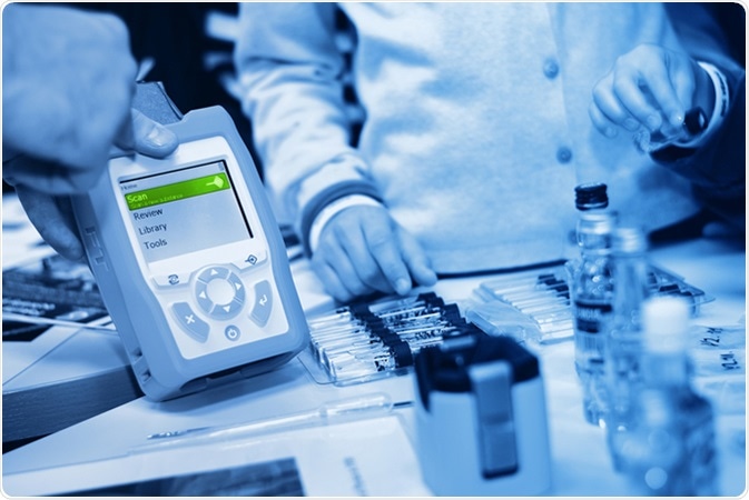
Portable Raman Spectrometer. Image Credit: Forance / Shutterstock
Principle
The basis of the technique is the Raman effect. This is based on the way the electronic cloud around a molecule interacts with an external electrical field produced by monochromatic light incident on the sample.
Depending on how easily the molecule can be polarized, a dipole moment is induced within the molecule. In other words, inelastic scattering of light from a laser or other monochromatic light source occurs in the spectrum between near-infrared to near-ultraviolet waves.
Here observed bands arise in the spectrum due to changes in the polarizability of the molecule in response to its interaction with light. Each band thus represents a specific molecular vibration and a particular energy transition.
These are plotted to provide a so-called molecular fingerprint which is unique for each molecule.
Procedure
A laser beam is used to illuminate the sample. A lens is used to collect the radiation from the illuminated spot, which is then directed into a monochromator. Any elastic scattered radiation, which results from the laser line (Rayleigh scattering), is removed by filtering. The remainder which is scattered inelastically is dispersed to fall on a detector.
Inelastic scattering refers to the change in wavelength and hence energy of the incident photon due to its interaction with the molecule, putting it into a different vibrational and rotational level. That is, if the final state of the molecule is a higher-energy than the initial state, the scattering results in a photon of lower energy to conserve the total system energy.
The opposite happens if the final state of the molecule is lower in energy than the initial. This results in a shift in frequency between the incoming and scattered photon, either a downshift or Stokes shift, or an upshift or anti-Stokes shift. The intensity of the shift is obviously dependent upon the rovibronic states of the molecule.
Inelastic scattering is weak compared to Rayleigh scattering, and therefore many instruments have been developed over time to separate the two, such as holographic gratings and photomultipliers were used as detectors. Currently notch or edge filters are the instrument of choice for laser rejection and CCD detectors.
Variants of this technique include surface-enhanced, resonance, tip-enhanced and stimulated Raman spectroscopy.
Advantages
The Raman effect has some benefits compared to infra-red spectroscopy such as:
- Avoiding the need for sample preparation
- Aqueous solutions can be used as water scatters light very weakly
- Ordinary glass sample holders are sufficient for use
- Carbon dioxide does not have to be purged as it is a weak scatterer
- The results reflect chemical structure accurately
- Both organic and inorganic molecules can be studied
Applications
Raman spectroscopy has become a widely used method of chemical analysis and molecular characterization of many compounds and chemicals.
It has become invaluable in solid-state physics, for the characterization of materials and delineation of the crystallographic orientation of studied specimens.
It is also proving its worth in nanotechnology, tissue imaging, and detection of pharmaceutical agents, including illegal drug detection, among numerous other uses.
Forensic Investigation Enhanced by Raman Spectroscopy Research at UAlbany
References
- http://www.inphotonics.com/raman.htm
- http://www.kosi.com/na_en/products/raman-spectroscopy/raman-technical-resources/raman-tutorial.php
- http://www.andor.com/learning-academy/raman-spectroscopy-an-introduction-to-raman-spectroscopy
- http://www.sciencedirect.com/science/article/pii/S2090536X15000477
- http://www.spectroscopynow.com/details/education/sepspec1882education/An-Introduction-to-Raman-Spectroscopy-Introduction-and-Basic-Principles.html?tzcheck=1,1,1,1,1,1,1,1,1,1,1&&tzcheck=1
- https://www.tu-darmstadt.de/
Further Reading