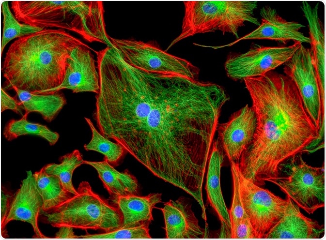
Cancer-associated fibroblasts (CAFs) are a major component of the cancer stroma. CAFs enhance the malignant features of nearby cancer cells, increasing the likelihood of metastasis.
P53 is a protein that exhibits tumor suppressive actions. It is well known that mutations in P53 can lead to cancer, but it is now also becoming apparent that alterations in P53 can lead to the formation of CAFs.
 Image Credit: Caleb Foster / Shuttterstock
The role of cancer-associated fibroblasts in cancer
Recent studies suggest that CAFs contribute to tumor proliferation, invasion, and metastasis. This is achieved through the secretion of various growth factors (GFs), cytokines, and chemokines, as well as through extracellular matrix (ECM) protein degradation.
Cancer cells that were cultured with CAFs had down-regulated E-cadherin expression compared to cancer cells that were cultured with normal fibroblasts. As E-cadherin is an epithelial adhesion molecule, this change allows the cancer cells cultured with CAF to migrate more easily, contributing to metastasis.
CAFs express matrix metalloproteinases (MMPs), such as MMP-2 and MMP-9. These MMPs degrade collagen and laminin, which subsequently causes the breakdown of the basement membrane. This contributes to cancer cell proliferation and invasion.
Angiogenesis is needed for the growth of malignant tumors. CAFs release various GFs and cytokines to induce angiogenesis. These include vascular endothelial growth factor (VEGF), platelet-derived growth factor (PDGF), stromal cell-derived factor 1 (SDF-1), fibroblast growth factor (FGF), and interleukin-6 (IL-6).
Origins of cancer-associated fibroblast
Past studies have reported that many different cells could be the origin of CAFs. Resident tissue fibroblasts, bone marrow-derived mesenchymal stem cells, and hematopoietic stem cells are examples of possible predecessors of CAFs. This makes CAFs heterogenous.
The conversion of CAFs from normal fibroblasts can occur in many ways. One method involves cancer cells reprogramming resident tissue fibroblasts to become CAFs, using small non-coding RNA molecules known as microRNAs or miRNAs (such as miR-31 and miR-214).
Reactive oxygen species (ROS) also promote the conversion of fibroblasts into highly migrating myofibroblasts via the accumulation of the hypoxia-inducible factor-1α (HIF-1α) transcription factor and the CXCL12 chemokine.
Finally, bone marrow-derived mesenchymal stem cells (BM-MSCs) can alter secreted transforming growth factor-β1 (TGF-β1), which promotes the formation of CAFs from fibroblasts.
Hematopoietic stem cells (HSCs) normally differentiate into several cell types from the myeloid and lymphoid lineage, including monocytes, erythrocytes, neutrophils, natural killer cells, and plasma cells. Alteration of differentiation factors can cause HSCs to differentiate into CAFs instead of normal HSC derived cells.
P53 and cancer-associated fibroblast generation
P53 is a tumor suppressor that blocks cell proliferation by causing the transcription of growth inhibitory genes (e.g. p21), but it can also tag a cell for apoptosis in the event of excessive DNA damage by causing the transcription of pro-apoptotic genes (e.g. Bax, Puma, Noxa).
Past studies have discovered that the expression and activity of p53 are decreased in CAFs. The loss of p53 allows the fibroblasts to overcome senescence, enhances expression of CAF effectors, and promotes stromal and cancer cell expansion. These changes show that p53 acts to protect against the formation of CAFs.
More recent research has discovered that the conversion of normal fibroblasts into CAFs involves the alteration of p53 from tumor-suppressive actions to tumor-supportive actions. The transcription targets of the CAF p53 are changed so it becomes a positive regulator of cancer-promoting genes and proteins.
The CAF p53 also upregulates features involved in tumor cell migration and invasion. This is an interesting discovery as it raises the possibility that tumor progression may involve the conversion of the role of the p53 gene.
CAFs as key components of malignancies
In conclusion, CAFs are a key component of some cancers. Their activity promotes proliferation, invasion, and metastasis of tumors. CAFs can be formed in many ways, which makes researching them a difficult task. P53 normally acts to guard against cancer formation, but it can be repressed or converted to perform tumor supporting actions which can aid in the formation of CAFs.
Acquiring more knowledge about the role and formation of CAFs (but also on the specifics of how p53 contributes to CAF formation) will undoubtedly provide a better understanding of tumor formation and progression. This information could allow for better treatment of tumors in the future.
Further Reading