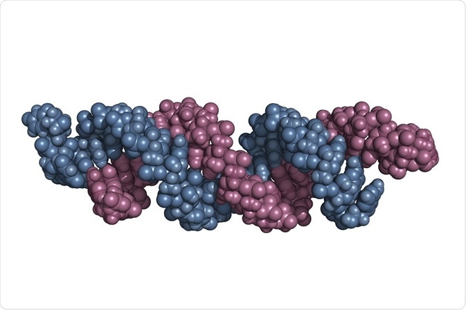
N6-methyladenosine (m6A) is the most commonly known RNA modification, and the methylation of adenosine at the nitrogen 6 position, which causes this. It is especially found in abundance in mRNA of eukaryotes and in tRNA, rRNA, snRNA, etc. A few studies have demonstrated that RNA m6A plays a vital biological role in the management of cellular metabolic activities.

Credit: Petarg/ Shutterstock.com
The methyltransferase enzymes, METTL3 and METTL14, help in “writing” of m6A RNA modification, and the modification can be reversed by FTO and ALKBH5 m6A demethylases. This promotes degradation and translational regulation of m6A-modified transcript and helps in determination of m6A modifying sites, which is important to study the possible biological role in RNA m6A modification at the transcriptome level. Several methods for identification of m6A modification have been developed to explore the change in functions of the protein.
m6A-seq and MeRIP-seq
Initially, two methods were developed, namely, m6A-seq and MeRIP-seq. These methods depend on the immunoprecipitation (IP) of disintegrated RNA fragments containing m6A sites, and followed by sequencing of these fragments. Both these techniques are defined as chromatin IP sequencing (ChIP-seq) and RNA-seq. The concept is similar for these two but MeRIP-seq differs from m6A-seq methodologically.
A whole RNA or mRNA is used in m6A-seq. First, a good quality of RNA sample is taken fragmented chemically using ZnCl2. A part of the fragmented sample is set as an input control. The resulting fragments are of size 100 nucleotides (nt). These fragments are exposed to IP process, where they are incubated with an m6A-specific antibody to bind with the m6A site. The unbound fragments are removed and the eluate and input control are sequenced in cDNA library and reads are obtained.
The reads are quality controlled and mapped to a reference genome by a short read aligner and mapped. The mapped reads are fed into peak calling algorithm called model-based analysis of CHIP-seq data (MACS). The m6A peaks are detected by comparing the input and the sample. The accuracy of the peaks is tested with multiple expectation maximization for motif elicitation (MEME), then the methylated motif are corrected with PeakAnnotator.
Single-nucleotide level methods
A method called SCARLET is used to detect the methylation of segments at m6A sites. Another method for detection of RNA modification is the technique that allows sequencing single molecule at real time, known as single-molecule real-time (SMRT).
PA-m6A-seq
The above developed methods (m6A-seq and MeRIP-seq) involve anti-m6A antibodies for binding and enhancing the m6A-containing RNA fragments, and detected the distribution of m6A through high-throughput sequencing methods in mammalian transcriptomes. As a result, it was shown that m6A levels are high at 3’-UTR around stop codons and within exons. As these methods include RNA fragments of size 200 nt, it is very hard to precisely locate the actual site of m6A modification.
The SCARLET approach was also established to accurately spot the methylation positions with single-nucleotide resolution, but it is very time consuming and not applicable for high-throughput applications.
PA-m6A-seq is a photo-crosslinking-assisted m6A sequencing approach, which can map m6A in a mammalian transcriptome with high precision. In this technique, photo simulative ribonucleosides (4-thiouridine or 4SU) are merged into mRNA. The oligo-dT-conjugated magnetic beads are used to separate 4SU inserted mRNAs. Then the full-length RNA is immunoprecipitated than the fragments and the sample is exposed to UV light of wavelength 365 nm. Later, the crosslinked RNAs are broken to about 30 nt using RNAse.
The Illumina TruSeq Small RNA Prep Kit is used to sequence the purified RNA. The crosslinking takes place only at a particular m6A sites which can be detected using RT-PCR. The authentication of the results from PA-m6A-seq was obtained by comparing and analyzing with higher-resolution map. The methylation sites pinpointed by PA-m6A-seq can be verified by SCARLET and m6A-seq/MeRIP-seq.
The higher-resolution map also indicates a clustering nature of m6A accumulation on transcripts, which is comparable to the cytosine methylation on genomic DNA. PA-m6A-seq labeled the numerous sites where methylation occurs in transcripts as MALAT1. In short, PA-m6A-seq technique is broadly practical to investigate exact m6A sites in different organisms and the UV-crosslinking approach is attuned with the study relating to nucleic acid and proteins.
M6A-LAIC-seq
Although the location analysis of transcriptome-wide m6A levels by m6A-seq (or MeRIP-seq) has led to significant understanding, methods to quantify the ratio of methylated to non-methylated transcripts (m6A level) on a transcriptome are lacking. An m6A-level and isoform-characterization sequencing technique (M6A-LAIC-seq) has been developed to measure the ratio of RNA distribution. This method sequences the intact full-length transcripts instead of fragmenting the RNA in addition to the quantification of m6A levels.
The study on the relationship between the number of m6A peaks and m6A levels showed that more the peaks found, the higher the m6A levels. About thousand extremely methylated genes depending on m6A levels were termed as transcription regulation and RNA metabolic process regulation. The m6A levels vary significantly between different cell types.
Further Reading