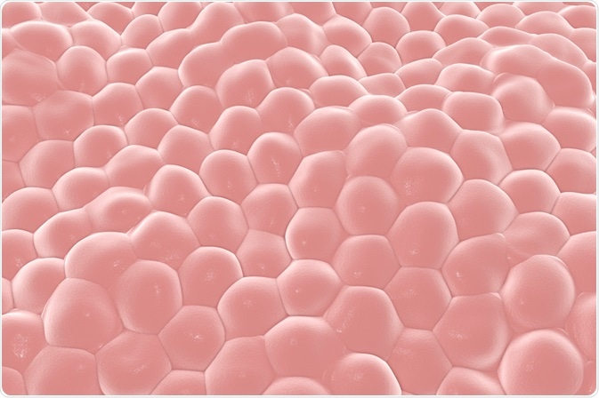
Human cells adhere to extracellular matrices, such as modified cell culture dish surfaces, and stretch into specific shapes by crawling along the solid support. But how strong does a surface need to be so that the cells do not slip?
Scientists from Japan have recently shown that a protein monolayer deposited on a liquid-liquid interface between water and a perfluorocarbon solvent is strong enough for human mesenchymal stem cells (hMSCs) to adhere and spread out.
 Kateryna Kon | Shutterstock
Human cells are naturally surrounded by extracellular matrices to which they bind via cell membrane proteins. In our bodies, a common extracellular matrix component is collagen, a fibrous protein that forms assemblies with other collagen chains.
Cells bind to specific components of the extracellular matrix via membrane-anchored proteins such as integrin that forms stable connections. On the cytoplasmic side, i.e. the cell’s interior, integrins can connect to components of the cytoskeleton, such as actin fibers which spread throughout the cytoplasm and stabilize cell shapes.
Thus, the interior skeleton can be anchored to stable, extracellular scaffolds, and the cell can move along or spread over the scaffold. This is necessary for cells to adopt their specific cell shape, such as an extended shape in the case of muscle cells.
In laboratory cell cultures, human cells are commonly grown on a stable solid surface which is modified with anchoring molecules, e.g. polylysine, or in 3-dimensional hydrogel cultures where the cells can connect to polymeric fibers that compose the gel network.
There is a great research interest in 3-dimensional cell cultures as they are more representative of natural tissues, where cells are surrounded by extracellular matrix and by other cells. However, the stiffness of the gel-polymer fibers needs to be optimized so that they can carry the tension of an anchored cell that intends to stretch itself into its desired shape.
Researchers from Japan, led by Professor Katsuhiko Ariga, have recently shown that a protein monolayer deposited at an interface between a perfluorocarbon and an aqueous liquid can be strong enough for cells to adhere and stretch, raising new possibilities for optimizing materials for cell culture.
Positioning Anchors at a Liquid-liquid Interface
The researchers tested two different perfluorocarbon solvents with low water solubility, to form an interface with an aqueous cell culture medium. The first was the more viscous perfluorodecalin (PFD) and the second the less-viscous, nitrogen-containing perfluorotributylamine (PFTBA).
The perfluorocarbon liquids were overlaid with aqueous protein solutions, and the proteins spontaneously formed monolayers at the interface. By testing with the protein BSA (bovine serum albumin), the investigators showed that a stronger protein monolayer formed at the PFTBA interface.
Subsequently, they prepared monolayers from the protein fibronectin, which can serve as an anchor for human mesenchymal stem cells (hMSC) by binding with their membrane protein integrin.
The scientists used microscopy to evaluate the binding of cells to the protein monolayer and to determine how much the cells can stretch by measuring their surface area.
In parallel, they analyzed the protein monolayer stiffness by AFM (atomic force microscopy) and the protein-folding in the monolayer by ATR-FTIR (attenuated total reflection – Fourier-transform infrared spectroscopy).
Denatured Protein Monolayers can Hold a Cell
When the perfluorocarbon compound PFTBA was used, strong protein monolayers formed, and ATR-FTIR showed that these were composed of denatured proteins. Since protein denaturation often lowers the water-solubility of the protein, networks can form by hydrophobic collapse.
The researchers could show that hMSCs could well adhere to these layers and adopted a stretched shape, demonstrating that the monolayers could withstand the tension applied by a cell while it is stretching.
The monolayers on the perfluorocarbon PFD meanwhile were more flexible, and the extend of protein denaturation was lower. As proteins in these monolayers could diffuse more easily, they did not serve as a strong anchor, and consequently, the hMSCs remained in a rounded shape rather than stretching along the monolayer surface.
This research could help to better understand how cells respond to surfaces with different stiffness, as the study authors demonstrated that the flexibility of the protein monolayers can fairly well be modified by changing the interface-forming liquids.
As the study authors highlight further, the knowledge that protein monolayers can supports cells to adopt their natural shape could lead to improved cell culture hydrogels. Irrespective of the gel-forming molecule, it could be considered to attach stable protein monolayers so that adhered cells do not slip when they apply a tension.
While it has not been tested in the present study whether the cells adhered to the interfacial protein monolayers would be able to grow and divide, it would be beneficial for future studies to know if longer-term experiments with growing cell cultures are possible as well.
Source
Jia X et al., Modulation of Mesenchymal Stem Cells Mechanosensing at Fluid Interfaces by Tailored Self-Assembled Protein Monolayers. Small 2019, 15 (5), 1804640; DOI: 10.1002/smll.201804640.
Further Reading