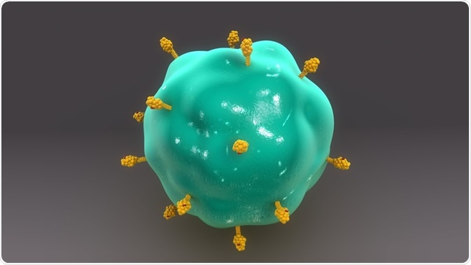
Flow cytometric immunophenotyping involves analyzing a heterogenous population of cells in order to identify a cell population of interest.
 Image Credit: sciencepics / Shutterstock
Immunophenotyping steps
- Cells of different lineages are identified and characterised as mature or immature cells.
- Abnormal cells are identified based on differences in antigen expression.
- Differences in normal and abnormal cells, such as the presence or absence of certain antigens are compared and documented.
- The data is then analyzed to make diagnosis (if needed), and additional tests such as immunohistochemistry, conventional cytogenetics, and fluorescence in situ hybridization (FISH) are carried out to confirm the diagnosis and make further conlusions.
- The data may also be used to develop prognostic tests for specific diseases.
Combining immunophenotyping with flow cytometry
Immunophenotyping can involve numerous cell types. The following sections deal with applications of immunophenotyping in flow cytometry for five kinds of samples.
Mature lymphoid neoplasms
Neoplasm or abnormal growth of cells of mature lymphoid tissue includes two kinds of lymphomas: chronic lymphoid leukemia and non-Hodgkin.
This group of neoplasms is characterized by an immunophenotype that is similar to normal lymphoid cells. They also do not have antigenic features associated with immaturity, including TdT, CD34, or CD45.
Mature lymphoid cells can be divided in to B cells, T cells, and natural killer (NK) cells. Studies have shown that a multicolor flow cytometry can be used to identify Hodgkin lymphoma in patients.
Mature B-cell lymphoid neoplasms
Immunophenotyping can be used to diagnose B-cell lymphoma, by identifying abnormal cellular antigens. This method can also be used to identify targets for therapy based on antibodies and also provide prognostic information regarding the expression of molecules, such as CD38, and ZAP-70, among others.
Two kinds of abnormalities in phenotype are used for identifying neoplastic mature B-cells: immunoglobulin light chain restriction and aberrant antigen expression. Mature B cells only express a single type of immunoglobulin light chain – kappa or lambda, whereas cancer cells express several other antigens.
Mature T and NK-cell lymphoid neoplasms
Immunophenotyping is also used to identify mature T or NK-cells. However, it is more difficult to identify the abnormal phenotypes of T or NK-cells using immunophenotyping compared to identifying abnormally shaped B-cells. This is because the classification of T and NK-cells is less established and involves input of information from several sources to confirm its identity.
After identifying a population as T or NK-cells, flow cytometry can be used to further detect the presence of markers, such as CD25 and CD52. The methods used to derive the immunophenotypic information are flow cytometry and paraffin section immunohistochemistry (IHC).
Plasma cell disorders
Plasma cell disorders involve increased serum or urine gamma globulins, and they can be divided in to reactive proliferation and monoclonal gamma pathology. Aberrant plasma cells are characterized by the following behaviors: increased number, abnormal phenotype, abnormal clonality, and a combination of morphology, radiology, and clinical findings.
To identify the abnormal plasma cells, and distinguish between lymphoid and plasma cells, flow cytometry is performed. Immunophenotypic data can also be used to derive information for prognosis.
Blastic neoplasms
Leukemia and lymphoma may also present with blasts and abnormal cells in the blood or body fluids and immunophenotyping again can be performed to identify immature or abnormal cells, and to distinguish them from immature cells that may be present in bone marrow and thymus.
Further Reading