Oxygen is an essential molecule for anaerobic organisms, acting as a terminal electron acceptor in the process of cellular energy generation. It is also used as a substrate in several enzymatic reactions. Oxygen homeostasis is important for the healthy maintenance of the entire organism.

Image Credit: Teeradej/Shutterstock.com
Oxygen concentration (OC) in cells can change dynamically due to imbalances in consumption and response to the microenvironment and cellular activity. For example, due to impaired vascular networks, the OC in tumor cells is lower than healthy cells, as the oxygen supply is limited. Pancreatic β cells increase OC for insulin secretion.
These are just a couple of factors amongst many that can lead to changes in intracellular oxygen consumption. To fully understand cellular oxygen dynamics, accurate assessment of the intracellular OC is therefore fundamentally important. Results of studies on intracellular OC dynamics are important for research into many medical conditions.
Experimental methods
Pathways that are involved in intracellular oxygen consumption are complex. Specialist techniques and equipment have to be employed to analyze and understand them. There are several different experimental approaches to studying tissue and cellular OC.
These include Microelectrodes, EPR (electron paramagnetic resonance), and optical methods.
Microelectrodes
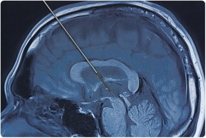
Using a microelectrode is a common method to measure oxygen consumption. A microelectrode is a biopotential electrode with an ultra-fine tapered tip. It is either inserted directly into a tissue, or an array of microelectrode needles can be placed against a tissue.
There are some drawbacks to the microelectrode method. A surface measurement can provide oxygen tension histograms over a wide field, but identification of the recording site is limited by the presence of the opaque electrode. Obtaining measurements at different sites in the field is difficult due to constant recalibration caused by the electrode having to be removed each time to take a new measurement.
Electron paramagnetic resonance spectroscopy
Electron paramagnetic resonance (EPR) is a type of spectroscopy. It was first observed by Y. Zavoisky in 1944 and has been studied widely since then.
It is analogous to NMR (nuclear magnetic resonance) spectroscopy, but rather than atomic nuclei, electron spins are excited. This method is extremely useful for detecting free radicals and reactive oxygen species (ROS.) ROS is the natural by-product of normal oxygen metabolism. Free radicals and ROS are short-lived and chemically distinct, making them difficult to detect in dynamic environments such as biological systems.
As reactive oxygen species have important functions in homeostasis and cell signaling, including being critical mediators in such processes like wound healing and angiogenesis, several research studies use EPR to study them.
The advantages of this method include high sensitivity, the ability to carry out repeat measurements, and low toxicity.
Optical methods
Optical methods are very good for studying oxygen concentration within tissues and cells as they rely on light, which is non-invasive and suitable for sequential monitoring. Methods include fluorescence resonance energy transfer (FRET) and two-photon imaging using luminescent quenching. Luminescent dyes are used to sense oxygen in cells.
When phosphorescent dyes such as PtTCPP are irradiated by light, grounded electrons are excited to a higher level, producing a photoexcited triplet state. This triplet state is quenched by oxygen, meaning that phosphorescence is highly sensitive to the oxygen concentration in the sample. This can be used to measure oxygen concentrations in real-time in a laboratory with minimal set-up.
Conclusion
As we can see, the oxygen concentration is an important marker of various biological processes and how it affects a wide range of conditions and diseases in the body is the subject of ongoing research, utilizing these methods and many others. Each method listed here has its advantages and disadvantages, and researchers should be well versed in the use of each of them.
Sources
Hyodo, F et al. (2010) In vivo measurement of tissue oxygen using electron paramagnetic resonance spectroscopy with oxygen-sensitive paramagnetic particle, lithium phthalocyanine, Methods Mol Biol. 610 pgs. 29-39.
https://www.ncbi.nlm.nih.gov/pubmed/20013170
Microelectrode – an overview, Sciencedirect.com
www.sciencedirect.com/.../microelectrode
Weiwei He et al. (2014) Electron spin resonance spectroscopy for the study of nanomaterial-mediated generation of reactive oxygen species, Journal of Food and Drug Analysis Vol. 22 Issue 1, pgs. 49-63
https://doi.org/10.1016/j.jfda.2014.01.004
Dmitriev, R. I and Papkovsky, D. B (2012) Optical probes and techniques for O2 measurement in live cells and tissue, Cellular and Molecular Life Studies Vol. 69 Issue 12, pgs. 2025-2039
https://doi.org/10.1007/s00018-011-0914-0