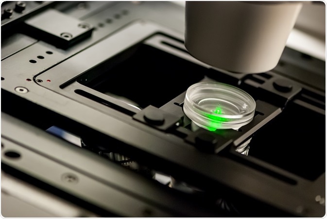
Fluorescence resonance energy transfer (FRET) is a mechanism of energy transfer that is widely utilized in biological experiments such as molecular structure examination. It is often referred to as a “spectroscopic ruler,” due to its nature wherein the distance between two fluorophores is determined as the ratio of fluorescence intensities.

Credit: Micha Weber/ Shutterstock.com
Application of FRET biological molecules is considered to be inevitable, as its measurements are highly sensitive at distances that are at nanometer scale. FRET measurement can be performed with the single molecule sensitivity alone, as it is based on fluorescent measurement technique.
Applications of FRET in DNA hybridization detection
Application of FRET in hybridization of DNA probes enhances the hybridization in two aspects.
- Signal from the probes that are unreacted is extinguished when hybridization is performed based on FRET. This avoids the need for washing procedures and solid support when hybridization reaction is observed in real-time as a homogeneous assay. Advantage of this homogeneous type of assay is its less complexity and speed, so that it can be easily adapted for automated performance.
- In vivo hybridizations can be directly performed in live cells by detecting only the probe signals that are hybridized and due to increased speed of hybridization in solution. FRET hybridization probes of different formats have been developed.
Applications of FRET in detection of DNA mutation
Energy transfer systems can either be created (TDI assays) or disrupted (invader assays) by oligonucleotide FRET probes that have been specifically developed for detection of DNA mutation. The invader assays use degradable probes like 5’ nuclease assays; moreover, they employ two degradable probes rather than one and one non-degradable probe working in concert.
Both linear and hairpin-shaped oligonucleotides are employed in developing branched DNA structures and then they are consequently destroyed during the reaction. The other advantage of this approach is that it results in effective signal amplification and does not require PCR for detecting very few target sequences. The distinct advantage of TDI assays is that FRET probes are produced in detection process but it requires PCR amplification.
Application of FRET in protein studies
Cell surface protein interaction plays a vital role in the transmembrane signaling process. The significant final outcomes of ligand receptor interactions are clustering of receptors and change in their conformation. FRET is used as a tool for identifying the distance relationship and supramolecular organization of cell surface molecules. In protein dynamics, protein folding is the most significant process that is widely investigated. smFRET has been utilized to learn more about folding and unfolding transition dynamics of two-state and larger multi-domain molecules.
Application of FRET in biological membrane mapping
Interaction of proteins that is present in the surface of the cell is essentially important for transmembrane signaling process. Clustering of the receptors and the variations in their conformations are the key factors that affect the final result of the interaction of ligand receptors. Cell surface supramolecular organization and determination of distance relationships can be efficiently performed by FRET.
Application of FRET in biological membrane mapping had resulted in lectin receptors clustering, cell surface distribution of receptor tyrosine kinases and hematopoietic clusters of differentiation molecules, and conformational variations of major histocompatibility complex I molecules on ligand binding and membrane potential change.
Applications in cell biology
FRET imaging that uses GFP spectral mutant adds the capability to monitor and locate ion binding and protein–protein interactions in living cells. The significant transmembrane receptor proteins that are involved in adhesion and cell signaling are often known as integrin.
For instance, in-vivo explicit interactions of intermolecular integrin are detected, which is enabled by FRET microscopy that is comprised of a pair of CFP/YFP FRETs. The local Rac–effector coupling is induced by guiding Rac to the membranes and detaching it from Rho-GDI.
Furthermore, due to its homogeneous dispersion in the cell, Rac is constitutively active; it interacts selectively with effectors at particular areas of the cell edge. The origination and termination of Ca2+ signaling in specific cellular sections such as endoplasmic reticulum, nucleus, or cytoplasm can be noticed by calculating the ratio change in the intensity of donor fluorescence and acceptor molecules present in the live cells.
FRET applications in other biomolecules
It has been thought that the enzymatic reactions that are driven by dynamics are conformational. An enzyme in an open structure is supposed to wait for a substrate and catalyze it in a closed structure. Then the reaction further progresses among the molecules in an asynchronous manner.
This implies that adenylate kinase (AK) has been in equilibrium between the closed and open structure despite the nature of the substrate binding, which alters only the rates of transition. In contrast to NMR or X-ray crystallography, standard approaches for determining the biomolecular structure, smFRET measurement does not need the molecule to attain a higher order structure.
Further Reading