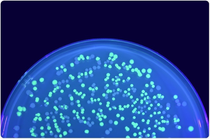
A fluorescent tag, commonly referred to as a label or probe, is a fluorophore that may be used to determine the distribution and quantity of a particular target biomolecule both in vitro and in vivo, in conjunction with fluorescence microscopy techniques.
 Image Credit: KPWangkanont / Shutterstock.com
The high degree of specificity gained through the use of proteins as targets or targeting agents has meant that fluorescent protein tags are abundant in the field of biotechnology. Three of the most common labeling methods are outlined below.
Intrinsically Fluorescent Proteins
Many proteins already present in the cell or tissue under examination are intrinsically fluorescent, and can be observed directly using fluorescence microscopy.
A fluorescent 25 kD protein harvested from the jellyfish Aequorea Victoria known as green fluorescent protein (GFP) has, however, become among the most employed protein fluorophores thanks to its versatility and adaptability, providing a string of engineered fluorophores with excitation and emission wavelengths ranging across the visible spectrum.
They are relatively compact and inert, forming a barrel-shaped tertiary structure of 11 externally-facing β-sheets shielding a helix that is responsible for the fluorescence.
These proteins can be bound to other proteins of interest at the N- or C-terminus without significantly affecting the function of these proteins. DNA recombinant technology even allows the GFP gene to be inserted into the DNA of a cell such that the GFP protein is expressed in conjunction with the protein of interest, indicating expression by the presence of fluorescence.
GFP is, however, comparatively large and sterically hindered compared with small-molecule fluorophores. They are frequently more sensitive to environmental changes such as fluctuations in temperature and pH, and generally produce a less intense signal that necessitates greater photon bombardment, inducing issues with cell viability and photobleaching.
Self-Tagging Proteins
In cases where intrinsically fluorescent proteins are unsuitable for the desired purpose, small molecule organic dyes may instead be used. In general, they tend to produce more intense fluorescence, be more stable, and provide a wider range of emission and excitation wavelengths.
In order to attach these probes to proteins in situ, in live cells, enzymes can be utilized that bind with the probe to a target peptide sequence. Specific enzymes optimized to purpose are commonly employed for this task, O6-alkylguanine-DNA alkytransferase (AGT) and its variants being among the most common.
O6-alkylguanine and O6-benzylguanine are molecules upon which this enzyme is active, and they can be bound with fluorophores that are incorporated into proteins via a cysteine amino acid on the protein of interest with the aid of AGT (marketed as SNAP tag). A modified form of the enzyme marketed as CLIP tag allows fluorophores bound with O6-benzylcytosine to be used instead,
Another enzyme, haloalkane dehalogenase, sourced from bacteria, is widely marketed as the HALO tag and operates in a similar manner. Being sourced from bacteria ensures that the enzyme is unlikely to interact significantly with biomolecules present in mammalian cells.
Covalent Bonding of Dyes
GFP, and the SNAP, CLIP, and HALO tags are each relatively large proteins that remain bound with the target protein of interest, potentially affecting the normal behavior of the target protein once bound. Instead, cell membrane penetrating biarsenical dyes that bind with peptides genetically introduced to the cell can be used. They are directly integrated into the target protein.
The peptide sequence is introduced into the target protein sequence and must follow the pattern Cysteine-Cysteine-X-X-Cysteine-Cysteine (X being any other amino acid) and be in a helical structure, which rarely occurs in nature.
Upon encountering this amino acid sequence the biarsenical dye forms a strong covalent bond with impressive affinity. This method can be used to examine protein dynamics in living cells, and also in vitro to label purified proteins.
The smaller molecular weight of the dyes involved in this process minimally affects the mobility of the target protein, and in addition, the twin covalent bonds formed between the fluorophore and protein prevent unrestricted rotational mobility between the two.
The larger size of protein fluorophores also limits their addition to the N- or C-termini of target proteins, while small organic molecules can penetrate the tertiary structure of the protein and label internally, allowing many more fluorophores to be present and thus producing a more intense signal.
Another dye, the membrane-permeable carboxyfluorescein succinimidyl ester, operates by bonding with lysine amino acid residues and other primary amines via the succinimidyl group, and can linger within cells for long periods.
Since it is relatively non-specific it is not ideal for detailing cell structure, but can be highly useful in tracking tagged cells over multiple generations, with the concentration of the dye being halved in each cell per generation.
Quantum dots represent a unique class of fluorophores made from nanometer-scale crystals of semiconductor metals. They produce highly intense fluorescence and are more resistant to photobleaching than small organic molecules, and the excitation and emission wavelengths can be finely tuned by material and size control.
A variety of targeting ligands can be attached to the quantum dot, including highly specific antibodies, peptide sequences, and nucleic acids. They can be introduced to living or fixed cells and allowed to distribute appropriately.
A number of long-term protein tracking experiments have taken advantage of the improved stability and half-life of quantum dots compared with both intrinsically fluorescent proteins and small-molecule fluorophores.
Source
Crivat, G. & Taraska, J. W. (2012) Imaging proteins inside cells with fluorescent tags. Trends in Biotechnology, 30(1), pp.8-16. https://www.ncbi.nlm.nih.gov/pmc/articles/PMC3246539/
Los, G.V. & Wood, K. (2007) The HaloTag™. In: Taylor D.L., Haskins J.R., Giuliano K.A. (eds) High Content Screening. Methods in Molecular Biology, vol 356. Humana Press. doi: 10.1385/1-59745-217-3:195 https://link.springer.com/protocol/10.1385%2F1-59745-217-3%3A195
Uttamapinant, C., White, K. A., Baruah, H., et al. (2010) A fluorophore ligase for site-specific protein labeling inside living cells. Proc Natl Acad Sci U S A, 107(24), pp.10914-10919. https://www.ncbi.nlm.nih.gov/pmc/articles/PMC2890758/
Griffin, B. A., Adams, S. R. & Tsien, R. Y. (1998) Specific Covalent Labeling of Recombinant Protein Molecules Inside Live Cells. Science, 281(5374), pp.269-272. https://science.sciencemag.org/content/281/5374/269.long
Griffin, B. A., Adams, S. R., Jones, J. & Tsien, R. Y. (2000) Fluorescent labeling of recombinant proteins in living cells with FlAsH. Methods in Enzymology, 327, pp.565-578. www.sciencedirect.com/science/article/pii/S0076687900273023?via%3Dihub
Sahoo, H. (2012) Fluorescent labeling techniques in biomolecules: a flashback. RSC Advances, 2, pp.7017-7029. pubs.rsc.org/en/content/articlelanding/2012/ra/c2ra20389h#!divAbstract
Werwie, M., Fehr, N., Xu, X., Basche, T. & Paulsen, H. (2014) Comparison of quantum dot-binding protein tags: affinity determination by ultracentrifugation and FRET. Biochimica et Biophysica Acta, 6, pp.1651-1656. https://pubmed.ncbi.nlm.nih.gov/24361618/
Further Reading