Hypertrophic cardiomyopathy is a genetic heart condition where the heart muscles become thickened and stiff, making it difficult for the heart to pump blood around the body effectively.
Usually the muscular wall of the left ventricle is affected, although the condition may also cause thickening of the muscle in the right ventricular wall. The thickened walls reduce the chamber’s capacity to hold blood during diastole and can cause reduced volume of pumping with each heartbeat. This leads to reduced blood flow from the heart. This in turn may cause symptoms such as dizziness, fainting, chest pain and shortness of breath.
Hypertrophic cardiomyopathy is inherited in an autosomal dominant pattern, meaning each child born to a parent with the condition has a 50% chance of inheriting it.
The condition is very common and affects about one in 500 people in the UK. Most people who have hypertrophic cardiomyopathy do not experience any symptoms and the condition may therefore go undiagnosed. For a minority, however, significant problems may develop including angina, lightheadedness, shortness of breath and arrhythmias that are life threatening, putting a person at risk of sudden cardiac arrest. Hypertrophic cardiomyopathy is the most common cause of sudden death in young people.
Diagnosis
Genetic testing
Hypertrophic cardiomyopathy is caused by the presence of at least one gene mutation that affects proteins present in the heart muscle. The cells of this tissue become disorganized rather than being arranged in straight lines, which may contribute to the arrhythmia that develops in some cases.
Since hypertrophic cardiomyopathy is inherited, people who have a first-degree relative with the condition are usually offered genetic screening to identify whether they have inherited a mutation and are at risk of the disease. However, genetic testing is not completely reliable, as little is understood about the underlying genetics. For this reason, mutations are only detectable in around half of families with the condition. The other tests doctors most commonly use to confirm a diagnosis of hypertrophic cardiomyopathy are described in more detail below.
Echocardiogram
This test enables a specialised observer to use ultrasound waves to visualize the heart’s pumping action and assess whether there is muscle thickening, obstructed blood flow or abnormal movement of the mitral valve. There are two forms of echocardiography, namely, transthoracic echocardiogram and transesophageal echocardiogram.
The first technique creates images of the heart using an ultrasound generator through a transducer placed on the chest skin. This form of echocardiogram is the technique doctors generally use to diagnose hypertrophic cardiomyopathy.
For the second technique, a tube is used to pass a transducer down into the esophagus where it can generate more detailed images of the heart. This may be advised if a standard echocardiogram fails to create images that are clear enough.
Electrocardiogram (ECG)
Here, the functioning of the heart’s electrical signaling system is analyzed using electrodes placed on the skin that measure impulses generated in the heart. This can tell whether the heart chambers and heart rhythm are normal or not.
Exercise test
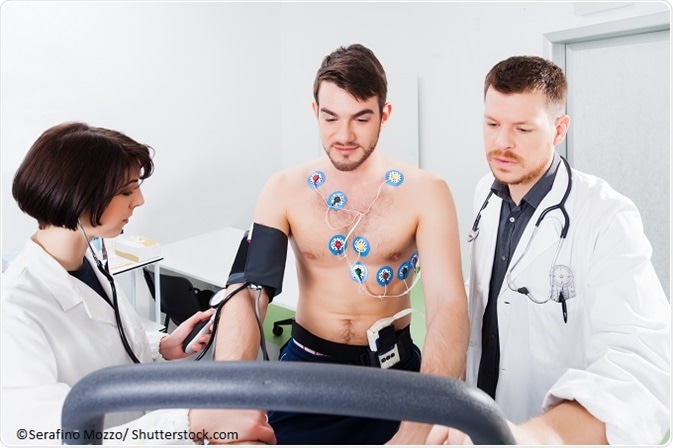
This involves the performance of continuous ECG monitoring while the individual is exercising on a treadmill, to determine how well the heart is functioning during periods of increasing activity. This will tell whether exercise triggers arrhythmia and how much exercise a patient can manage before symptoms arise. Sometimes the test is carried out in conjunction with echocardiography.

Magnetic resonance imaging (MRI)
A cardiac MRI scan may be performed, which generates detailed heart images using magnetic fields and radio waves.
Holter heart monitor
Here, a portable ECG device is fixed around the neck or waist. It records the heart’s electrical activity continuously over the course of one or two days.
Cardiac catheterization
This involves the use of a catheter inserted into a blood vessel and passed through to the heart chambers, to release dye into the bloodstream, followed by X-rays. These images are then used to evaluate the heart and blood vessels.
References
- https://www.cardiomyopathy.org/
- https://www.heart.org/HEARTORG/Conditions/More/Cardiomyopathy/Hypertrophic-Cardiomyopathy_UCM_444317_Article.jsp
- https://www.bhf.org.uk/
- http://www.mayoclinic.org/diseases-conditions/hypertrophic-cardiomyopathy/home/ovc-20122102
Further Reading