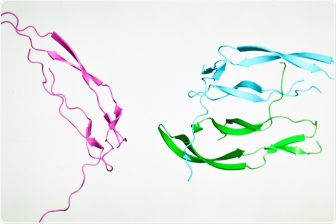
The three-dimensional structure adopted by biological macromolecules largely determines their role in different cellular processes. Several different imaging techniques ranging from X-ray crystallography to cryo-electron microscopy have been successfully applied for the structural characterization of numerous macromolecules.
 Image Credit: Sergei Drozd / Shutterstock.com
What are macromolecules?
Although up to 80% of living matter is made up of small molecules including inorganic ions, organic molecules and, primarily, water, the remainder of living matter is comprised of macromolecules. Macromolecules can be in the form of proteins, polysaccharides or genetic material like deoxyribonucleic acid (DNA).
Proteins, for example, are the most abundant type of cellular macromolecule present within cells, as they typically account for approximately 20% of the cell’s total weight. Many of the proteins present within cells are enzymes, which are responsible for catalyzing important biochemical reactions that provide energy and nutrients to the cell. In addition to enzymes, other protein functions assist in the overall motility of cells, maintain cellular rigidity, and support the transportation of molecules across cell membranes.
Advanced cellular imaging methods
Recent progress in the development of high-resolution cellular imaging modalities have equipped scientists with the ability to investigate biological processes as they occur in their natural environment.
As these imaging methods continue to advance, researchers are not only interested in identifying the role of macromolecules in different cellular reactions, but also expanding current knowledge on the structure of these macromolecules.
In fact, understanding the structure of macromolecules is central to understanding their function, as many molecules, particularly enzymes, will adopt complicated three-dimensional (3D) structures to catalyze a specific chemical reaction.
Traditionally, the structure of macromolecules has been studied through either X-ray crystallography or nuclear magnetic resonance (NMR) spectroscopy). Although traditional X-ray imaging is insufficient to study individual protein complexes, X-ray crystallography can provide important structural information on proteins when multiple copies of these macromolecules are arranged together in a 3D crystal.
Similarly, NMR, which measures distance-dependent interactions between atoms, can also be used to infer the structure of certain macromolecules to determine their dynamics and behavior during interactions with other molecules.
Several other imaging methods have been used for macromolecular structure determination, some of which include transmission electron microscopy (TEM), single particle analysis, 3D reconstruction, cryo-electron microscopy (cryo-EM) and electron cryotomography (cro-ET).
Determining macromolecule structure with electron microscopy techniques
It is estimated that proteins scatter electrons at a significantly stronger rate that is up to ten thousand times greater compared to their X-ray scattering patterns. The electrons that are scattered from these macromolecules can be accelerated in certain electrical fields to provide information on the distance that is present between the atoms that make up these macromolecules, thereby allowing for their structure to be precisely calculated.
Although previous applications of electron microscopy (EM) techniques for macromolecule structure characterization were modest compared to those utilizing X-ray crystallography and NMR, recent progress in EM techniques have transformed structural biology studies.
Compared to traditional EM techniques that destroy protein samples after imaging, cryo-EM, which allows samples to be kept frozen at cryogenic temperatures, can protect proteins from the destructive effects of radiation damage while also preserving their structure when exposed to the high vacuum of EM systems.
A typical cryo-EM imaging of a protein sample will require several microliters of the sample to be applied onto a thin film of amorphous carbon material that is placed on top of a metal grid.
After any excess fluid is blotted away, the metal grid is placed into liquid ethane to produce a thin layer of vitreous ice to allow for several copies of the protein to arrange themselves into different orientations, all the while maintaining the ultrastructure of the specimen.
Through the use of an electron microscope operating at liquid nitrogen temperature, multiple two-dimensional (2D) images of the protein sample are then obtained through the holes that are present within the amorphous carbon film. These images, taken together, will then be used to create a high-resolution single 3D construction of the protein.
After the images are combined, the macromolecular structure can be determined through a method known as sub-tomogram averaging, which combines repeating 3D densities of the specimen inside reconstructed volumes known as tomograms.
Future outlook
Advanced imaging technologies like cryo-EM and cryo-ET have met the growing demand to visualize minute details of macromolecules that are present within cells.
When applied to bacterial specimens, for example, such technologies can provide structural information on the bacterial macromolecules that are responsible for antimicrobial resistance and bacterial pathogenicity.
This type of information not only improves the fundamental understanding of the behavior and activities of these microorganisms but can also be used during the design of therapeutic strategies aimed towards their elimination and/or circumventing antimicrobial resistance.
References and Further Reading
Melia, C. E., & Bharat, T. A. M. (2018). Locating macromolecules and determining structures inside bacterial cells using electron cryotomography. Biochimica et Biophysica Act (BBA) – Proteins and Proteomics 1866(9); 973-981. doi:10.1016/j.bbapap.2018.06.003.
Lodish, H., Berk, A., Zipursky, S.L., et al. (2000). Section 1.2, The Molecules of Life in: Molecular Cell Biology 4th ed. New York: W. H. Freeman. Available from: https://www.ncbi.nlm.nih.gov/.
Ruprecht, J., & Nield, J. (2001). Determining the structure of biological macromolecules by transmission electron microscopy, single particle analysis and 3D reconstruction. Progress in Biophysics and Molecular Biology 75(3); 121-164. doi:10/1016/S0079-6107(01)00004-9.
Fernandez-Leiro, R., & Scheres, S. H. W. (2016). Unravelling the structures of biological macromolecules by cryo-EM. Nature 537(7620): 339-346. doi:10.1038/nature19948.
Further Reading