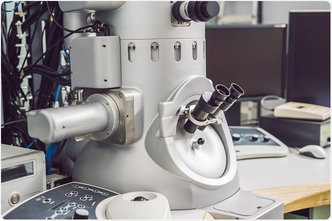
The term “cryo-electron microscopy” (abbreviated as “cryo-EM”) is used in a large number of experimental methods. It works on the principle of imaging the radiation-sensitive samples by a (TEM) under low temperature conditions.

Credit: Elizaveta Galitckaia/ Shutterstock.com
This technique is mainly aimed at the development of new approaches to enhance the image resolution of protein structures within cellular conditions. Attaining a maximum number of good-quality images is time-consuming in this technique; hence, this is still subject to further research into new ways of allowing the microscope to logically select quality imaging areas and eliminate poor quality images. It should also evaluate the image quality significantly in real time.
Cryo-EM—three-dimensional technique
In the three-dimensional (3D) cryo-electron tomography technique, tomography (a visual record produced by tomography) is considered as one of the ways to achieve the 3D design of macromolecular groups by utilizing electron microscopy.
In this technique, collecting a group of images is achieved with every image taken at various angles in relation with the incident electron beam direction. For acquiring images of thicker samples, such as large viruses, an imaging filter is used. This filter removes electrons that are inelastically scattered and helps to improve the contrast.
For procuring more information from the tomographic data, two types of strategies are followed:
- Averaging methods: This is used when a clear identification of definite molecular entities within the cells or outside the microorganism is done, or when a molecular group is imaged in vitro.
- Comparing multiple tomograms: To analyze morphologically varied organelles or cellular substructures.
Although the microscope is able to operate at 300 kV, the samples are required to be thin enough (<0.5 microns) to transfer the incident electron beam.
Two-dimensional technique in single particle analysis
The single-particle analysis is considered as a typically utilized variant of cryo-EM. In this technique, the structure of 3D reconstruction is generated by a combination of data taken from 2D presentation images obtained at various orientations. Here, similar copies from the protein complex are featured together to reconstruct the 3D structure.
Similar to other applications of cryo-EM in single particle imaging, the first step is the spread of complexes that are soluble through a hole in a carbon film. Later, the specimen is plunge-frozen, which creates a vitreous ice thin layer in order that it contains a group of the same copies at different orientations.
Starting from the fields of images, which consist of more molecular groups, the selection of a single particle is done manually or by an automated algorithm. Then statistical techniques such as principal component analysis, covariance analysis, or multivariate analysis can be done based on the difference in the structural appearance to sort the images.
While imaging the icosahedral viruses, it is found that by utilizing a single particle, two high resolutions are achieved: (1) Due to the presence of higher symmetry, it allows to gain the advantage of averaging among every virion. (2) The virions with a maximum size provide good contrast for perfectly analyzing the orientation of particle over image presentation.
Cryo-EM of ordered assemblies
The information regarding 2D images may be of higher resolution, but in a few cases it exceeds 3Å. By principle, near-atomic resolution can be achieved even without symmetry for structures extracted by the cryo-EM method. In addition, there is a high limitation in achieving the goal for unique macromolecular assemblies of intrinsic conformational heterogeneity. This challenge can be overcome by finding the experimental conditions that cause helical formation of the protein sample.
This type of strategy is highly effective in membrane proteins where 2D crystals are formed over the membrane plane. In some cases of bacteriorhodopsin, the approach of 3Å is allowed by structural determination.
New technical advances
In the past, the drawbacks in the cryo-EM field have been the significant time consumption and required complete concentration toward the work, which have stacked the progress of its wide use. In imaging with modern microscopes, the alignments and positioning are strongly fixed, and data collection is generally performed once for every multi-day period. However, it is an important challenge for compensating and maintaining the sample placement, especially in tomography and single-particle analysis.
At the mechanized stage, the samples’ orientation and position are determined accurately by using a micrometer. As the data acquisition is adjusted during this stage, the movement seems to have small changes repeatedly in sample coordinates. Using the collaboration of image shift and beam shift and defocus, they can be compensated. From a single hole, collection of various adjacent images can be performed by utilizing beam shift and image shift.
Owing to the working process of cryo-EM and the steps of automation and improvements, they not only increase the speed greatly and are easy to use but also have improved the data quality. The process of converting 2D images into 3D reconstruction involves image processing and specific computational power.