To communicate with its neighbors, animal cells use gap junctions which are symplasmic connections through which molecules can diffuse from one cell to another.
In higher plants, the communication occurs in part through plasmodesmata, which are bridge-like structures linking the cytoplasm of adjacent cells. Fungi primarily operate through pores which facilitate transport of molecules and form integral parts of development and function.

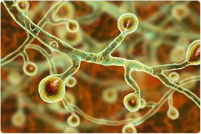
Blastomyces dermatitidis fungi, the causative agent of the disease blastomycosis affecting lungs, more rarely skin, bones, other organs, 3D illustration - Image Credit: Kateryna Kon / Shutterstock
Fungal Cell Wall
Filamentous fungi form a mycelium in which organelles and cytoplasm are distributed like a network. The fungal cell walls – septa – are often perforated by pores through which molecules and even larger organelles such as mitochondria can pass. These septal structures can either be uniperforate (one pore) or multiperforate (several pores), and different structures can be present within one organism.
Plasmodesmata
In 1966, a plasmodesmata-like structure was found in newly formed fungal cell walls. The resemblance to plant plasmodesmata was due to intracellular strands running through the pores from one cytoplasm to the other, resembling the desmotubule in plasmodesmata. More reports suggest that these strands can be associated with the endoplasmic reticulum (ER) or with desmotubules. The plasmodesmata-like pores are much narrower than “normal” pores and have been shown in the lower fungi. These lower fungi lack septa, so the plasmodesmata-like structures are present in “cross walls” which separate reproductive structures from vegetative cells.
Cytoplasmic Sharing
In higher fungi, the perforated septa through which fungi achieve direct cell to cell communication are present in hyphal tips. These pores support the colony growth but can also be detrimental. If one compartment becomes damaged, there is risk of leakage to all connected cells. To prevent this, the pores can be plugged. Similar to plasmodesmata in plants, the pores can be plugged by depositing proteins or organelles. An example of this is a group of membrane bound organelles called Woronin bodies which are normally located around the pore opening but move into the pore to plug it during an injury. In more ancestral fungi, the Woronin bodies are closer to the pore, whereas in other fungi they are located more distally to widen the pore and promote cytoplasmic flow.
Septal Pore Caps (SPCs)
In higher fungi the septal pores can instead be associated with septal pore caps (SPCs) due to the pores being more complex. At its base, the SPC is connected to the ER, and organelles can move through the pores. However, the nuclei are unable to pass through these pores except during specific times of development. This lends support to the theory that the septal pores have an important role in development in filamentous fungi and not just in limiting damage and cytoplasmic bleeding.
Cell-Cell Communication During Development
Type and Complexity of Pores
Variations in cell-cell communication can be introduced by type and complexity of pores which can differ within one organism. During development of sexual structures, pores are often more restricting than they are in the hyphae. The movement of organelles is restricted, whereas ions and small molecules can move freely.
Location of Pores
Cell-cell communication can also be influenced by the type and location of pore occlusions which also differs in the fruiting bodies. Recent evidence suggests that the septum in the fruiting bodies helps in the differentiation of cell types. A basidiomycete fungus lacking a specific cap protein also lacked SPC, resulting in stunted mushroom formation. Thus, the pore structure leads to selective transport, which helps drive cell differentiation during development.
Further Reading