Flow cytometry is a popular technique used to measure the expression of molecules, identify cells, and analyze cell size and volume. It most commonly involves labelling cells with fluorescent antibodies or ligands, which bind specific targets in the cell to be detected.
 Image Credit: Hakat / Shutterstock
Types of samples
The simplest samples to prepare for flow cytometry are suspended culture cells, waterborne microorganisms, bacteria and yeast. Whole blood is also relatively easy to use, because the red blood cells can be removed by lysis and the white blood cell types can then be identified.
Single cells need to be produced from solid tissues for flow cytometry. This is done by disaggregating the tissue. Mechanical disaggregation works well for tissues that are loosely bound, such as adherent cells from tissue culture, bone marrow, and lymphoid tissue.
In mechanical disaggregation, chopped tissue in a suspension is moved through a fine-gauged needle repeatedly. This can be followed by grinding, if necessary.
Tissue can also be disaggregated by enzymes. In enzymatic disaggregation, enzymes disturb interactions between proteins and the extracellular matrix that is holding the cells together. Enzymes are chosen based on factors such as pH, temperature, and other cofactors, which all influence the action of the enzymes.
Direct and indirect immunofluorescence staining
There are different staining techniques for flow cytometry. Among these is immunofluorescence staining, which can be direct or indirect. Direct immunofluorescence staining involves treating cells with a fluorescently labelled antibody.
Indirect staining is sometimes called the sandwich technique, wherein the primary reagent is not labeled but is instead detected by a secondary reagent that is fluorochrome-labeled. These can both be antibodies, where the secondary antibody has specificity for the primary one.
Overall, immunofluorescence staining is easy, but the binding specificity and intensity can be influenced by several factors.
Single cell staining
Single cell staining is a technique used for the detection of surface antigens. Lymphoid tissue or peripheral blood are examples of what single cell suspensions can be made from.
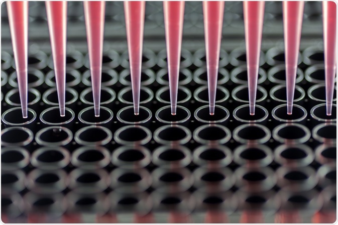
In this method, cells are incubated in tubes or microtiter plates (flat plates with many wells that can be used to house solutions) with antibodies. The antibodies can be unlabeled or labeled with fluorescence. The cells are then washed with a buffer solution to remove antibodies that did not bind to the cells.
Single cell suspension may also be used for the detection of intracellular antigens. This requires the plasma membrane to allow dyes or antibodies through, by fixed and permeabilized single cell suspensions. It can accurately measure cellular DNA content. Further, cell loss is low, which means it can be performed on just a few cells.
Protocols to prepare cells for single-cell suspension flow cytometry can be modified to detect antigens, which differ slightly in their sensitivity to denaturation, or in how the antibody stays bound to the antigen after permeabilization of the cell.
The internal and surface antigens of a cell can be measured simultaneously by staining both the cell surface for antigen expression, as well as the intracellular for antigen expression. However, the type of antibody labeling is important, as some, such as peridinin chlorophyll protein (PerCP) fluorescence, is lost after permeabilization.
Non-viable cell staining
Non-viable cells are dead cells, and have therefore lost their membrane integrity. This means they cannot be used in fixed or permeabilized cell preparations. 7-aminoactinomycin D, or 7-AAD, can be used here. Fluorescent 7-AAD can bind DNA, but this can be blocked by including actinomycin D, or AD, which is not fluorescent.
Tissue culture cells
Preparing cells from tissue culture is generally easier than single cell suspensions. The cells that are grown in suspension can be gradually poured into a centrifuge tube, centrifuged, and resuspended in a flow cytometry staining buffer.
Adherent cell lines need to first be detached. Once detached, remaining clumps need to be disassociated. Then they can be centrifuged and resuspended in a flow cytometry staining buffer and used for flow cytometry.
Further Reading