Bending Light
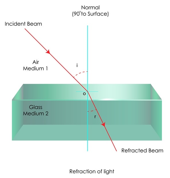
Since ancient times, it has been known that glass can bend light. Light is made up of electromagnetic waves and with properties such as wavelength and frequency, light usually behaves like a wave and can be diffracted.

Refraction of light in glass. Image Copyright: snapgalleria / Shutterstock
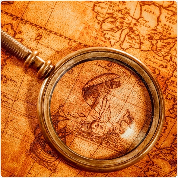
Once glass had been invented, the Romans started experimenting with it and testing it. They found that if the glass had a thick middle but was thin at the edges, objects would look larger when this “lens” was held over them. These early forms of the lens were referred to as magnifiers.

Image Copyright: Andrey Armyagov / Shutterstock
Early Light Microscopes Developed
During the 1590s, two Dutch spectacle producers further experimented with these early lenses. Zaccharias Janssen and his father Hans Janssen realised that if you put a small object in a tube containing several lenses, the object would appear very large when at the end of the tube and was much more enlarged than when a simple magnifying glass was used.
The pair only achieved a magnification of 9x and these early microscopes were more novelties than scientific instruments.
In the late 17th Century, Anthony von Leeuwenhoek from Holland invented a single lens, hand-held microscope that could achieve a magnification of 270x.
Using this lens, he went on to develop the first microscope that could actually be made use of. Leeuwenhoek found he was able to see structures that noone had seen before such as blood cells and bacteria.
In the same century, Englishman Robert Hooke was acknowledged as having discovered the smallest most basic unit of an organism – the cell.
He was also recognised as the first person to use a microscope with three lenses, the configuration used in today’s microscopes.
Microscopes go into Large Scale Production
There were few further developments made to the microscope until the middle of the 19th century, when sophisticated microscopes such as the ones we use today were developed. The German company Zeiss started to manufacture these refined devices.
During these early years, scientists worked to solve various problems with the microscopes, such as the unequal bending of light that hits the lens in different places. In 1830, Joseph Lister, realized that placing the lenses at specific distances from one another resolved this problem for all but the first in the series of lenses.
For the first lens, use of a low power lens with low curvature minimized this unequal light bending to the extent that the problem was almost eliminated.
Phase-Contrast Microscope Developed
Frits Xernicke developed the phase-contrast microscope in 1932. This device enabled researchers to study transparent biological materials.
Electron Microscope Appears
The use of visible light in microscopy limits the resolution that could be achieved, but this was problem was overcome in 1931 when two German scientists Max Knoll and Ernst Ruska discovered that beams of electrons could be used instead of light. The electron microscope could to be used to observe objects that were not visible using light microscopes.
Scientists working for corporations competed to develop the first commercial electron microscope and Ernst Ruska, working for Siemens, eventually achieved this in 1938. By the late 1930s, microscopes had been developed that could achieve resolutions as low as 10nm and by the mid 1940s, resolutions as low as 2nm had been achieved.
The main competitors in Europe were Siemens, Philips and Carl Zeiss. In the late 1930s, the scientists in Japan formed the Japan Electron Optics Laboratory that eventually manufactured the greatest variety of electron microscopes among all of the companies.
The early versions of electron microscopes used transmission electron microscopy. The first scanning electron microscope hit the market in 1965, which revolutionized the world of material science.
References
Further Reading