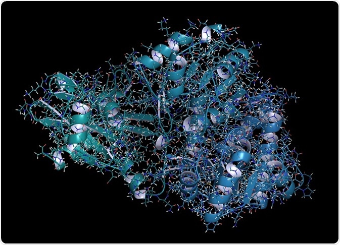
Bioluminescence resonance energy transfer (BRET) is a noninvasive, advanced cell-based assay technology used for detection of protein–protein interactions. BRET involves fluorescent and bioluminescent protein tags with corresponding emission and excitation properties for investigation of resonance energy transfer between the protein tags in their vicinity owing to their interactions.

© molekuul_be/Shutterstock.com
Identification of these protein–protein interactions provides vital information about various diseases and improves methods of treatment.
Working principle
The working principle of BRET is based on the Förster resonance energy transfer, which states that the efficiency of the transfer of energy between a donor and an acceptor is inversely proportionate to the sixth power of the distance of separation between them.
The bioluminescent donor usually opted for the BRET assay is Renilla Luciferase (RLuc), which is often separated from the sea pansy, Renilla reniformis. It enhances the oxidation of its substrate, coelenterazine, in order to obtain a blue light. The obtained emission spectrum overlies the excitation spectra of yellow fluorescent protein (YFP).
Procedures to set up a BRET assay
Setting up a BRET assay for examination of protein–protein interactions includes the following steps:
- Sorting and creation of donor/acceptor and substrate couple. The creation of the two proteins naturally bonded with either donor luciferase protein or acceptor fluorescent protein is dependent on the type of BRET.
- Designing and authentication of the relevant receptor along with appropriate controls. Determination of the most suitable protein is done because insertion of a negative/positive control is more essential to finalize the interaction specificity.
- Sorting of the cell system and co-expression of the two BRET bonded proteins at a relevant physiological level of expression.
- Detection of the BRET signal and fabrication of appropriate assay. The signals are measured from the member cells, cell suspension, subcellular sections, purified proteins, and also from culture medium.
- Evaluation of the BRET signal. The BRET signal is the ratio of the light signal of the acceptor emission relative to that of the donor emission. Then the net BRET value is computed by deducting the BRET background ratio from the BRET ratio acquired from the cells. The evaluation of acceptor by donor is done by dividing the prior fluorescence magnitude by the luminescence magnitude.
BRET derivatives
There are three different BRET derivatives that are widely used for laboratory purposes: BRET1, BRET2, and eBRET.
BRET 2 uses DeepBlueC substrate, which has extraordinary donor and acceptor emission separation capability, allowing the differentiation of donor and acceptor emissions easily and reducing the background noise signals. Due to low quantum yield and quick decay of the substrate, large numbers of cells are required for attaining high luminescence levels for detection of BRET. It also requires a high-sensitive apparatus.
eBRET has the advantage of high throughput screening and does not require frequent addition of substrates. eBRET should be performed only by using live cells at room temperature. Since it is dependent on the endogenous esterases, it cannot involve in the process of assaying cell extracts or purified proteins.
Applications of BRET
There are several applications of BRET which include:
BRET system for measuring dynamic protein–protein interactions in bacteria
The detection of dynamic protein–protein interactions taking place inside bacteria can be carried out using BRET technique with luciferase LuxAB. This is done to acquire information about the bacterial activities such as development and pathogenicity.
In the process, eYFP (enhanced yellow fluorescence protein) accepts irradiation from the LuxAB bacteria and produces yellow fluorescence. The occurrence of BRET is observed only when LuxAB and eYFP are bonded to the interacting protein pair FlgM and FliA. Further, the sirolimus-induced interaction between FKBP12 and FRB and the dose-dependent abolishment of the interaction by FK506 leads to the formation of FKBP12 ligand.
Application of BRET to clock proteins
BRET techniques can be used to explain the formation of homodimers by the interaction of clock proteins KaiA, KaiB, and KaiC encoded by circadian (daily) clock genes from cyanobacteria.
Application of BRET in imaging of mammalian cells
BRET imaging techniques are broadly used for analysis of protein interactions and several other methods for illustration of bacterial cultures, plant tissues, and mammalian cell suspensions. An EB-CCD camera combined with a micro-imager is used for imaging of BRET signals at the tissue, cellular, and subcellular levels.
The process of BRET imaging is used for supervision of protein interaction of COP1 in plant tissues that absorb various light spectra. Quantitative BRET magnitude can be obtained by introducing a rectifier of RLUC-EYFP emission profiles. In addition, the interaction of CCAAT/enhancer binding protein α (C/EBPa) in the nuclei of separated mammalian cells can also be detected using the BRET technique.
Other applications of bimolecular resonance energy transfer (BRET) are
- GPCR-β arrestin assay for monitoring G-protein coupled receptor activity
- Receptor dimerization
- cAMP detection
- Tyrosine kinase receptor activation
- Kinase activity
- Ca2+ detection
- Apoptosis assay
- Protease activity
Further Reading