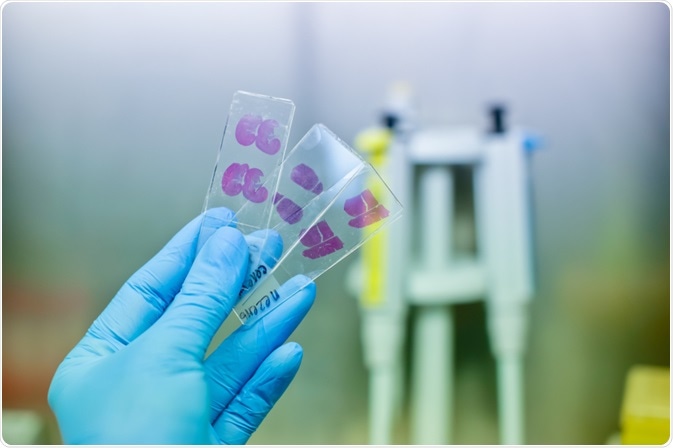
Histopathology is the study of how tissues change during disease. It is the gold standard for disease diagnosis, and advances in artificial intelligence will only increase the accuracy of this technique.

Vshivkova | Shutterstock
What is artificial intelligence (AI)?
Artificial intelligence is an ‘umbrella’ term encompassing both machine learning and deep learning, and is based on the idea that the machine can provide an optimal solution to a problem by passing information through levels of neural networks.
The major types of artificial intelligence (AI) learn by being presented with a labelled data set. In the case of histopathology, the AI algorithm will be exposed to diseased and healthy samples, from which it will pick up some distinguishing markers by itself.
How can AI be used in histopathology?
In a histopathology laboratory, a single diagnosis typically involves skilled specialists who have spent 11 or more years of training to correctly identify diseased tissue through microscopes. Pathological evaluation involves both diagnosis of the tissue, as well as prognostic assessment based on the tissue architecture and morphology of cells.
Initially, technological advances were limited to quality assurance and certain research applications. Hence, the development of AI tools that can accurately diagnose diseases is a huge advancement.
Artificial intelligence can be applied to detect and count cells, for example in mitotic events which may be relevant for cancerous cells. It can also be applied to detect segmentation and classify tissues as healthy or diseased. A key benefit with such artificial technology is that it can be applied to whole slide imaging, where the AI can automatically identify patterns in a whole slide.
Current applications of AI in histopathology
Current AIs have been shown to be capable of detecting breast cancer metastasis in a lymph node. Initial research with this has revealed that the AI is capable of achieving success rates comparable to a human specialist, with a specificity of almost 90% and sensitivity of 75%. The detected tumors measured 0.2 mm or were made up of low number of cells (i.e. less than 200 cells).
AI may also be particularly valuable for repetitive tasks, such as image analysis. Repetitive and tedious tasks tend to make human interpreters bored, which leads to higher error rates.
Will AI replace histopathologists?
Despite some early and rather exaggerated claims, histopathologists are unlikely to be replaced by AI. However, the development of AI will hopefully continue in order to increase efficiency and accuracy in diagnosis by carrying out preliminary diagnosis and quality assurance. The role of histopathologists will be to ensure that the AI are functioning as they should, and not misidentifying cell states.
Some have likened the shift to the drastic change in role and job duties that geneticists and bioinformaticians experienced with the advent of next-generation sequencing (NGS). However, others argue that the mere question of whether or not professional histopathologists will be replaced is like comparing water and fire – they are two very dissimilar things.
AI is based in high-level computing, whereas humans are high-level cognition, meaning they will reason a cognitive opinion based on training and experience with the influence of bias to produce the diagnosis. This allows patient-specific information to be accounted for, primarily to increase diagnostic value.
Potential complications
With pervasive use of AI in histopathology, it may be difficult to determine the correct magnification for the tissue analysis, ensuring correct annotation the training data sets should have and ensuring the suitability of the training dataset.
The training data set is of paramount importance, because AI falls under the “garbage in, garbage out” category. More specifically, if the data set is not constructed of information-rich examples, or if any annotations are incorrect, it can seriously impact the accuracy and ability of the AI.